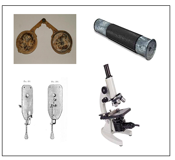
Day 1
Advance Preparation: Conserve time by setting up a microscope and three slides with “unknown” objects (e.g., table salt, insect leg or wing, onion cells) at lab stations before class. Also, write instructions for the microscope lab on chart paper:
- Take a microscope from the microscope station.
- Grip it according to your Microscope Lab Procedure.
- Carefully bring it to your station/area.
- Set it up.
- Insert the provided microscope slide.
- Accurately bring the view into focus.
- Raise your hand once you are finished in order to have your focus and set up checked.
- Once checked, return the microscope to its proper location and ask the next member from your group to repeat the steps.
Start the lesson by having students look at a few different structures under the microscope. Instruct students to try and guess what structure or part of a structure they’re looking at.
Group students according to the number of microscopes. Provide each group with three unknown microscope slides to look at. Ideas for unknown objects include table salt, a portion of an insect leg or wing, and a very thin slice of an onion layer. For better results, cut the onion with a razor blade to create the thinnest layer possible to allow the light to penetrate the onionskin.
Say, “There are unknown objects on each of the three microscope slides. Your objective is to figure out what you’re looking at during the three minutes given to you for each slide. Write your answers down on a piece of scratch paper. Raise your hand once you have come up with a guess, and I will come by to check your focus and insert the next slide for your group. You will all get a chance to operate the microscopes independently after the next section of the lesson, in which you’ll learn key microscope structures and handling procedures.”
Allow three minutes for each slide so all group members get a chance to examine the slide. Students will raise their hands between slides to have you change the slide out and focus the next slide into view. Once all groups have had a chance to examine each of the three slides, list students’ guesses on the board and review the answers with them as well as addressing any questions.
Hand out the Microscope Diagram (S-6-4-3_Microscope Diagram and KEY.doc) to all students and give them a few minutes to look over the structures. Hold up a real microscope and show them each of the parts. Have them label each of the parts on the diagram as you point them out on the microscope. Then, discuss the answers to the questions on the worksheet and have students record the answers. It is important that students understand how microscopes can extend human capabilities and enhance our understanding of the natural world.. Tell them, “At the end of the unit, you will have to demonstrate how to handle and use a microscope correctly as part of your unit assessment.”
“Now we are going to review the history of the microscope and learn how to use and handle it so you can operate one independently during the lab.”
- Display or hand out copies of the Microscope Timeline (S-6-4-3_Microscope Timeline.doc). Review the timeline of inventions and discoveries that lead to the modern compound microscope found in classrooms across the world. Elaborate, if needed, on the Timeline of inventions and their impact on science:1595: Zacharias Jansen built the first microscope.
- 1600s: Anton van Leeuwenhoek improves the microscope to 200x magnification.
- 1665: Robert Hooke used the first compound microscope to view thinly sliced cork cells.
- 1673: Anton van Leeuwenhoek first described living cells as seen through a simple microscope.
- 1838: Mathias Schleiden identified the first plant cells and concluded that all plants are made of cells.
- 1839: Thomas Schwann made the same conclusion about animal cells.
- 1858: Rudolf Virchow concluded that all cells come from already existing cells.

Source:http://nobelprize.org/educational/physics/microscopes/timeline/index.html
Explain the difference between a “simple microscope,” such as the one used by van Leeuwenhoek, and a “compound microscope,” such as those in the classroom. Simple microscopes just have one lens, while compound microscopes have multiple lenses, allowing for much greater magnification.
Address any student questions before handing out the Microscope Lab Procedure (S-6-4-3_Microscope Lab Procedure.doc). Instruct students to read through their copy of the Microscope Lab Procedure along with you as you model proper microscope handling and use. Call on student volunteers to read steps. Once the procedures are read, have students get into the same groups from the unknown object activity earlier in the lesson.
Display the chart paper with instructions that you prepared in advance. Tell students, “All groups will take turns sending one member at a time to follow the directions written on the chart paper. You may refer to your Microscope Lab Procedures during this exercise.” You can place any object in the microscope slide as long as the focus can be determined.
Provide feedback throughout the exercise to encourage proper handling of the microscope and its features. Once finished, wrap up the lesson by allowing students a few minutes to review the microscope parts using their diagrams. This will help them study for the end-of-unit assessment.
Day 2: Pond Water Lab
Prior to the lab, collect local pond/lake water or order it from a biological supply company (see Related Resources). Student groups will need about an ounce of pond water at the most.
Review the types of living organisms students will be searching for in the pond water by handing out to each student a Living Things Concept Map (S-6-4-3_Living Things Concept Map.doc) and displaying the Protista Pictures (S-6-4-3_Protista Pictures.doc) on an overhead or handing out copies. The concept map shows all six living kingdoms, highlighting the organisms students will be searching for in the lab, which are the protists. Students will learn more about Kingdom Protista in the Pond Water Lab Resources document (S-6-4-3_Pond Water Lab Resources.doc).
Group students according to the number of microscopes and hand out the resources document. Have students read through the document for a few minutes before setting up for the lab. Students from each group will complete the following tasks for the set-up procedure:
- One member from each group will collect the microscope.
- One student will collect the pond water and plastic pipette.
- One student will hand out the Pond Water Lab Sheets (S-6-4-3_Pond Water Lab Sheet and KEY.doc) and the Pond Water Resources handouts (S-6-4-3_Pond Water Lab Resources.doc).
Students will take turns operating the microscope until they have brought an organism into focus, allowing other group members to look at the organism and fill out their lab sheet accordingly. The next student in the group will then take over the microscope to search for the next organism. Specific directions are provided on the Pond Water Lab Sheets.
Once they have finished with the lab, have students answer the questions on page five of their lab sheets and collect them. Have students complete the take-down procedures. They may refer to their Microscope Lab Procedure sheets if needed to take down and handle the microscopes appropriately.
Extension:
- Students who require more practice with the standards can pair up with students who better understand the concepts to help them through the lab procedures. Students can also be given an appropriately modified lab procedure with fewer steps and more opportunities to answer in visual depictions, as opposed to short-answer requirements.
- As you model microscope handling and use, have students point to each part on the microscope at their station and say the parts aloud.
- Students who may be going beyond the standards can research other microorganisms found in typical pond water. Students may consult the library or Internet to do so. Have them make a poster that displays one of the organisms and general information about its classification, appearance, habitat, and habit (see Related Resources, Virtual Pond Water Web site).
